loading
What we do

Glaucoma, commonly known as ‘the silent thief of sight, is one of the most serious eye conditions, often progressing without noticeable symptoms until significant vision loss occurs. Recognizing its severity, we are committed to early and precise diagnosis using the most advanced equipment, and we offer the latest and most effective treatment solutions to protect and preserve vision.

Non-contact type tonometer - Air puff tonometer.

PRODUCT INTRODCTION

Portable non-contact tonometer, It is an innovative product that measures Intraocular pressure with a simple manipulation by patients themselves, at home or office without the risk of anesthesia, pain, or contamination. Our device is easy to use by anyone

Standalone non contact tonometer with Corneal thickness measurement Auto soft air puff , Auto alignment , Auto focus, Auto switch OS&OD , Print report auto paper cutting , Electric chin rest.

REBOUND TONOMETER

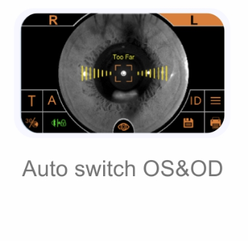
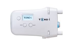
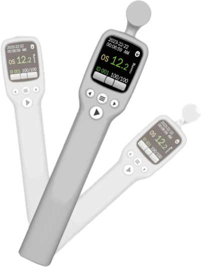
Handheld measurement, easy to operate. Automatic positional alignment with

guidance and intuitive user interface. Can be operated by both right and left handers, no specialised skills required,just load, align and measure.

No epidural anaesthesia, no chemicals, fast and accurate measurements, high patient comfort. 200° of positional freedom, regardless of whether the patient

is in the standing, sitting, elevated or supine position.

Trabecular Meshwork Cutter

Excise the trabecular meshwork in ophthalmic surgery, restoring the natural aqueous humor outflow pathway. It can be performed as a standalone procedure or combined with cataract surgery, effectively reducing IOP and improving the patient’s quality of life

Goniosynechialysis Spatula

Primarily used for goniosynechialysis; it can also be used for the precise localization of angle foreign bodies and cyclodialysis clefts.

The angle structures can be directly observed through specular reflection without the aid of an agonioscope.

During angle separation, gentle pressure applied to the iris root allows simultaneous observation of the angle and separation of adherent iris tissue. No microscope tilting or patientnhead position adjustment is needed during use